Neuropsychologists: How They Help Care For Your Brain
Answer a few questions and we'll provide you with a list of primary care providers that best fit your needs.
Clinical neuropsychologists know their way around your brain. They’re specialists within the field of clinical psychology who know how the brain works, how to assess your brain functions, how brain disorders impact them, and how to design effective treatments, such as brain tumor surgery, to protect your brain’s operation.
“A neuropsychologist is the type of doctor who specializes in brain functions – things like memory, attention, concentration, spatial skills. Anything that your brain can do, a neuropsychologist can measure,” says Fadi Tayim, Ph.D.,
clinical neuropsychologist and director the Brain Mapping Center of Premier Health’s Clinical Neuroscience Institute.
The Functions Of the Brain’s Lobes
The various parts, or lobes, of the brain, control different functions.
“Most often people come and see me because they have some type of frontal lobe impairment,” Dr. Tayim says. “They say, ‘I'm not functioning the way that I used to and I want to figure out what's wrong.’"
The frontal lobes, right behind your forehead, control executive, or higher order, brain functions. Among these, he says, are “planning, organizing, multitasking, even eliminating distractions in your environment, these are all things that are moderated and mediated by the frontal lobes.”
Difficulty with memory is another common concern. “The temporal lobes (situated behind your ears) are really our memory center. Memory is stored all over the brain, but when we talk about forming new memories, retrieving memories, and consolidating memories, that's really the temporal lobes and that's done in the hippocampus.”
The hippocampus, at the edge of each temporal lobe, is one of the first parts of the brain to suffer damage from Alzheimer’s disease and other forms of dementia.
Other parts of the brain include the occipital lobes, at the back of the brain, which are responsible for visual perception, and the parietal lobes, at the back and top of the head, which help make sense of what you see.
“The temporal lobe, parietal lobe, and occipital lobe work together,” Dr. Tayim says. “It's very rare for one part of your brain to function independent of the others, because I can see an object, but without the other lobe involvement, I can't make sense of what I'm seeing.”
How Neuropsychologists Assess Brain Functions
Neuropsychologists like Dr. Tayim see patients with a variety of disease processes that affect the brain, such as Parkinson's disease, Alzheimer's, head injuries, epilepsy, and brain tumors.
To assess the impact of disease processes on the brain, neuropsychologists use a variety of tests, depending on the disease. For instance, for Alzheimer's type dementia, tests are selected to test for memory.
Tests may include puzzles, paper-and-pencil tests, computer tests, and fine motor and refined skills testing, Dr. Tayim says.
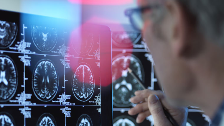
Mapping the Brain
Neuropsychologists also use functional magnetic resonance imaging (fMRI) to assess and map brain function. “By giving a patient tasks to perform in the scanner, in the MRI, we're able to see blood flow, oxygenated blood flow, to determine whether that region of the brain is properly functioning towards that activity that we're asking them to do.”
Dr. Tayim adds that testing is done prior to neurosurgery, including brain tumor surgery, “because the neurosurgeon, typically, has concerns as to whether the area of the brain that is affected by a tumor, epilepsy, or any part of the brain that is to be resected (surgically cut out), if that resection will result in some kind of debilitating impairment.”
He adds, “Using fMRI we perform a series of tasks. I will have a patient move their finger so I can see how the left or the right side of the brain, what we call ‘lights up’ when they are performing that task.
“Another task that we can perform is asking a patient to read a sentence or listen to speech so that we can determine how, for example, the posterior temporal lobe, the receptive language area, is functioning. Any area that is activated typically should not be resected. That's the reason why we do functional MRIs for a patient leading up to their surgery.”
Answer a few questions and we'll provide you with a list of primary care providers that best fit your needs.
Source: Fadi Tayim, Ph.D., Clinical Neuroscience Institute; American Academy of Clinical Neuropsychology; American Psychological Association; National Institutes of Health