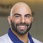
Rachel’s Story: New Awake Craniotomy Procedure Allows Local Mom to Return to Her Active Life
Rachel Borger is always on the go. The 39-year-old mother of two from Dayton is constantly in motion, from work to her children’s sports practices
“It’s a hectic life,” says Rachel. “I’m always busy running the kids around.”Rachel’s active lifestyle of being a mom hit a speed bump after years of experiencing what she believed were panic attacks and brief periods of blindness in her left eye
“It happened after I gave birth to my daughter,” explains Rachel. “One day, I was at work and my coworkers called the medics during one of my episodes. If it weren’t for them, I probably would have let it go forever.”
After an MRI evaluation eight years ago, doctors discovered that Rachel had a lesion on the part of her brain’s left temporal lobe that is responsible for vocabulary, word-retrieval, and recalling the names of famous places and landmarks.
Rachel was on medication and visited her neurologist regularly. However, a routine MRI in 2020 found that the lesion on her brain had grown.
Rachel was found to be a candidate for the new awake craniotomy procedure at Miami Valley Hospital – a procedure performed by Ania G. Pollack, MD, neurosurgeon at the Clinical Neuroscience Institute (CNSI), and Fadi M. Tayim, Ph.D., neuropsychologist and director of the CNSI Brain Mapping Center, the first in the Midwest.
“Rachel was the perfect case,” says Dr. Pollack. “She had a benign vascular lesion in the eloquent part of the brain that was getting bigger. Rachel had developed intractable seizures requiring more and more medications. Seizures and increasing doses of antiseizure medications changed her lifestyle and overall functional status.”
Awake craniotomies are frequently — but not always — used for primary brain tumors such as gliomas (including glioblastoma, astrocytomas, and oligodendrogliomas) as well as metastatic disease. Awake craniotomy is typically performed for cases where critical brain functions may be affected, like speech, motor skills, and memory. Patients who participate in an awake craniotomy also must feel comfortable with the idea of waking up during surgery, though they won’t remember it happening.
“When Dr. Pollack told me I was going to be awake, at first, I was a little nervous,” remembers Rachel. “However, I did my homework and watched plenty of videos to see that this was actually something that could help me even more.”
“With this procedure, the brain is exposed, and we wake the patient,” explains Dr. Pollack. “We have special protocols to keep the patient comfortable. We wake up the patient and stimulate the brain prior to any tissue removal to avoid potential deficits after surgery.”
Stimulating parts of the brain to avoid deficits is referred to as cortical stimulation mapping, or brain mapping, a key component of the awake craniotomy procedure. Brain mapping is vital to understanding the brain’s geography, anatomy, and how each area functions, and allows for a highly individualized surgical plan and greater precision during surgery. While Dr. Pollack stimulates the brain, Dr. Tayim evaluates the functions that are specific to each area using the NeuroMapper intraoperative platform. For Rachel, the focus primarily was to evaluate core language skills and naming ability.
Dr. Tayim says that it’s not only removing the lesion that’s difficult, but also getting to it sometimes. “You have to go through, or bypass, very important cortical functional brain tissue, and navigate that without causing any new deficits for a person,” he says. “Awake craniotomy, or intraoperative mapping, allowed me to guide the surgeons through tricky areas of Rachel’s brain, helping them make decisions during surgery that would protect her from greater deficits after surgery,” Dr. Tayim says.
Dr. Pollack, along with Daniel Gaudin, MD, Ph.D., FACS, a fellowship trained neurosurgeon at Miami Valley Hospital, removed the mass on Rachel’s brain, as well as a part of the brain that had been causing additional seizures.
Rachel participated in two surgeries. The first surgery was the actual resection of the lesion and preparation for the seizure surgery; the second surgery was solely dedicated to her seizures.
The second procedure took place 10 days following the first, and Rachel remained in Miami Valley Hospital afterward for observation.
“I felt completely fine and was able to walk around and do some tasks on my own,” explains Rachel. “It took a little getting used to, but overall, it wasn’t as bad as I thought.”
Just a few months after her surgeries, Rachel says that her vision problems and other symptoms disappeared.
“It’s great to see the patient before and after surgery,” Dr. Tayim says. “Rachel has her smile back, her life back, she’s participating in activities with her kids. It’s so good to see her being able to do that.”
“It's like I never had surgery,” says Rachel. “It's so weird. There are so many people who have no idea I even had brain surgery, and when they found out they were like, “What?’”
Rachel cites the awake craniotomy for giving her the ability to experience activities she hasn’t been able to enjoy in years.
“I’m back to working out and going almost every day during my lunch breaks,” says Rachel. “This has given me back so much and I no longer experience excruciating pain in my head anymore.”